3D mammography catches breast cancer sooner, reduces false results
May 15, 2019
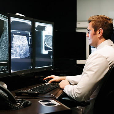
Q & A with Radiologist Christopher Henley, M.D., fellowship trained in breast and women’s imaging
Studies have shown that 3D is better than 2D mammography at detecting breast cancer. In the fall of 2018, the NCH Breast Center made 3D mammography the standard of care for all patients. Dr. Henley explains how this difference is improving patient outcomes.
What is 2D mammography and how does it work?
2D mammography remains the standard of care in most hospitals for breast imaging. The breast is placed in compression and two standard views are taken of both breasts. One view images the breast from top to bottom and one from side to side. The images are a summation of all the breast tissue imaged within the plane. The images are then reviewed by a radiologist to evaluate subtle changes from prior years.
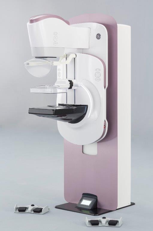
How does 3D mammography work?
3D mammography is performed in conjunction with the 2D mammogram. The X-ray arm sweeps in an arc over the breast, taking multiple images. Instead of the single image taken with a 2D view, we get approximately 100 or more images per view. This creates a more complete picture of the breast, as we are able to evaluate the breast tissue on a millimeter-by-millimeter scale instead of the summation of the 2D images alone. Essentially, we’re able to look inside the breast tissue instead of just an overview of the breast. We’re better able to evaluate the shapes, sizes and effects of surrounding tissue on any suspicious abnormality.
Does 3D reduce the number of false positives?
Yes. It decreases the recall rate (having to get called back for additional scans) because we’re better able to differentiate a small breast cancer from benign overlapping breast tissue.
Why did NCH decide to use 3D mammography as its standard?
We’re able to detect smaller breast cancers that could be hidden; it allows us to decrease the callback rate; we’re better able to characterize suspicious lesions; and we have greater accuracy in determining if a breast needs a biopsy or additional testing.
What have the results been since switching from 2D to 3D?
3D mammography research has been found to increase the cancer detection rate by 30 to 40 percent. NCH data confirms similar findings.
What does this mean for patients?
If we’re able to find a cancer at an earlier stage, the patient has more treatment options available. This, overall, improves survival and reduces complications from breast cancer.
Does the 3D technology make mammograms any more comfortable?
The 3D technique is similar to 2D imaging in terms of positioning and patient comfort.
If something suspicious is detected, what happens next?
Once an abnormality has been identified on a screening exam, the patient will be brought back for additional diagnostic imaging. That could be an additional mammogram, an ultrasound or sometimes both. Generally about 8 to 12 percent of women will get recalled every year. Of that, about 15 to 20 percent of women will go on to have a biopsy.
Besides 3D mammography, what other advanced technologies are available for detecting breast cancer at NCH?
ABUS, (Automated Breast Ultrasound Screening) is offered as a supplemental screening tool. Much like 3D mammography, ABUS allows us to look through dense breast tissue. Some masses that may not be detectable on mammography may be better visualized with ultrasound. Since dense breast tissue is a risk factor for developing breast cancer, 3D mammography with possible supplemental screening with ultrasound on an annual basis is important.
The award-winning NCH Breast Center offers four imaging locations in Arlington Heights, Buffalo Grove and Schaumburg with short wait times and same-day results. Call 847-618-3700 to schedule your 3D mammogram today. Learn more about the NCH Breast Center at nch.org/breast.