Automated Breast Ultrasound Screening
We offer Automated Breast Ultrasound, or ABUS 3D screening—the only technology developed and approved by the FDA for screening women with dense breast tissue.
Only a radiologist reviewing your mammogram can make the determination that your breasts are dense. Forty percent of women in the U.S. have dense breast tissue, making them more likely to develop breast cancer.
If you have had a recent mammogram, ABUS may be the next screening exam for you. The advanced system produces a 3D image that allows a radiologist to “peer through” dense breast tissue and see areas that were not clearly visible on your mammogram.
ABUS screenings do not replace regular mammograms. Mammograms are still essential.
Advanced Diagnostics
If a potential problem is detected during your mammogram or ABUS screening, we will perform additional testing and give you the results quickly. We want to reduce your waiting―and worrying―time. We use the latest diagnostic procedures to provide accurate results.
Available procedures include:
- Targeted breast ultrasound—Our doctors use ultrasound imaging to determine if a detected mass is fluid-filled or solid before an exploratory biopsy is done.
- Core needle biopsies—If needed, samples of tissue from the breast can be safely obtained using a core needle biopsy. This is often the preferred biopsy method because it’s less invasive than surgery. Core needle biopsies can be completed using various guidance technologies. We can complete them using X-ray (stereotactic), ultrasound or MRI, depending on what your radiologist recommends.
- Intact breast excisional biopsies—Our surgeons use this traditional method to remove all or part of the lump.
Expert Care Close to Home
If even the smallest cancer is detected, our team of experts will work with you to create a treatment plan. Your team will include medical oncologists, radiologists, surgeons, radiation oncologists and a nurse navigator—all highly skilled and dedicated to ensuring you get the most comprehensive care.
A multidisciplinary clinic takes place each week to review results and treatment options for newly diagnosed breast cancer patients to minimize the time between diagnosis and treatment. The clinic team develops a personalized treatment plan.
Our multidisciplinary approach and quality care have merited numerous awards and accreditations, including:
- Three-Year Approval with Commendation by the Commission on Cancer of the American College of Surgeons
- American College of Radiology Breast Imaging Center of Excellence
- NAPBC – National Accreditation Program for Breast Centers
- Magnet® Status for Nursing Excellence—achieved by only eight percent of hospitals in the nation
Understanding Breast Density and ABUS Screenings
What is breast density?
Breast density is not a measure of how the breasts feel, but rather how the breasts look on a mammogram. It compares the area of breast and connective tissue seen on a mammogram to the area of fat. Breast and connective tissue are denser than fat and this difference shows up on a mammogram.
- High breast density means there is a greater amount of connective tissue compared to fat.
- Low breast density means there is a greater amount of fat compared to connective tissue.
Dense breasts are usually found in younger women. As women get older, their breasts become less dense and after menopause, breast tissue has a larger percentage of fat.
Why does it matter?
Breast cancer is more common in women with dense breasts and can be more difficult to detect with mammography. The denseness of the fibrous and glandular tissue can mask abnormalities in a mammogram.
How do I know if I have dense breasts?
Breast density is determined after your mammogram is read by the radiologist. Dense breast tissue is common; it’s found in 40 percent of women. If a radiologist determines that you have dense breast tissue, he or she will inform you and your doctor.
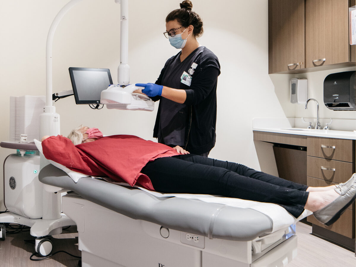
What happens during an ABUS screening?
The process is simple: You will lie down on the exam table and a layer of lotion will be applied to your breasts. Then a sheer membrane covering a transducer device is firmly positioned on your breast. The scanning process takes less than 60 seconds. ABUS screenings use sound waves (not radiation) to create state-of-the-art 3D ultrasound images of the breast tissue. After the screening, the radiologist can view the images along with your mammogram results to ensure every portion of the breast is thoroughly examined.
Is it painful?
You will feel some pressure as the transducer is positioned on your breast, but it is a pain-free process.
Do I still need a mammogram?
Yes. Mammography is still essential because (1) it finds more cancer cells, and (2) it detects calcifications in the ducts, which can be an early sign of cancer. ABUS screenings do not show calcifications. An ABUS screening is a secondary screening only for women whose breast density could be masking other abnormalities.
Schedule Now
To schedule a mammogram and possible ABUS screening, call 847-618-3700.